

Atypical Mole
Have you been told you have a precancerous or atypical mole?
Or maybe you were not satisfied with your doctor’s response to “What is a precancerous mole? What does atypical mean?”
Maria M. Tsoukas, MD, PhD, Assistant Professor, Dermatology Section, University of Chicago, will explain in detail just what a precancerous (atypical) mole means.
First of all, if you’re concerned about a precancerous or “atypical” mole, make sure that you consult with a dermatologist as opposed to your primary care physician.
Dr. Tsoukas explains, “Moles are common skin growths. They are correctly called melanocytic naevi (American spelling ‘nevi’), as they are due to a proliferation of the pigment cells, melanocytes.
“If they are brown or black in color, they may also be called pigmented nevi. Moles are benign in nature (harmless), but malignant melanoma (cancerous) may arise within a mole.”
About 30 percent of melanomas arise in a pre-existing lesion.
Dr. Tsoukas continues, “Some of the nevi appear since birth. There can be nevi that normally appear till around the 35th year of age.
“In pigmented and non-pigmented melanocytic nevi, we may identify clinically and histologically [with a microscope] certain atypical features.”
Atypia means the cells show some unusual features.
“In clinical examination by an expert, even when atypia is detected, malignancy cannot be excluded,” says Dr. Tsoukas.
“In those cases we obtain a specimen for histologic examination under the microscope or biopsy.”
Even if you’re planning on having a normal looking spot removed for cosmetic reasons, request a biopsy just in case.
Dr. Tsoukas says that in clinical examination, dysplastic (atypical) nevi are often bigger than other moles, and tend to be abnormally shaped and colored.
They can be anywhere including non-sun exposed skin.
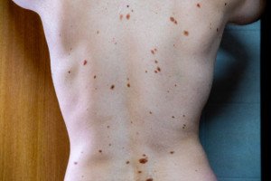
Atypical moles (dysplastic nevi). Shutterstock/Mikel Ugarte Gil
Atypical mole. Source: cancer.gov
When nevi are present with a lot of clinically defined atypical features, a biopsy is indicated to evaluate for melanoma — which kills one American every hour.
“In microscopic evaluation there are certain features that are met so as a tumor is defined as malignant,” explains Dr. Tsoukas.
“Dysplastic (atypical) nevi may present with some of those abnormal features under a microscopic evaluation.
“However, they do not meet all criteria to be diagnosed as melanomas under the microscope.”
Think of a continuum: At one end is benign, and at the other end is melanoma.
An atypical (dysplastic) mole sits somewhere between both ends of the continuum.
“This means that atypical nevi have some features that we see in melanoma, in clinical exam and under the microscope, but not all of them.”
So, in theory, any nevus that looks pathologically atypical can evolve into melanoma.
However, if the microscope shows mild atypia, this isn’t nearly as concerning as severe atypia. Moderate atypia falls somewhere in between.
Now, what percentage of melanomas actually arise from an atypical mole?
Dr. Tsoukas says those who have dysplastic nevi without family history of melanoma face a 7 to 27 times higher risk for developing melanoma.
However, this doesn’t mean that an atypical mole will necessarily transform into melanoma, although a severely atypical mole — as just mentioned — has the highest chance for melanoma transformation.
“We carefully examine and thereafter remove atypical moles (entire lesion) via biopsy or surgical excision techniques in the office.”
Having many dysplastic nevi is one of the risk factors for melanoma.
“Someone with many dysplastic nevi and personal history or with 1st-degree relatives who have had melanoma has an extremely higher lifetime risk of developing melanoma, than general population” says Dr. Tsoukas.
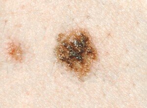
Melanoma under magnification. Shutterstock/Nasekomoe
Additional risk factors include: fair skin, sunburn history, indoor tanning, advancing age (though teens and children can get melanoma) and immunosuppression.
Remember the mnemonics for mole assessment: ABCDE
A – Asymmetry; one half of the mole does not match the other.
B – Border; ragged, notched or blurred.
C – Color; non-uniform and changing; mottled appearance; may include areas of red, white and blue; beware of new blackness in a mole.
D – Diameter; though melanoma is usually bigger than 6 mm (larger than pencil eraser) upon diagnosis, they can be smaller.
E – Evolving; beware of a changing mole and don’t hesitate to seek medical advice.
 clinical interests include diagnosis and management of patients at high risk for skin cancer, cutaneous oncology, laser surgery and aesthetic dermatology.


